A Giant Primary Intracranial Hydatid Cyst
Authors
Abstract
Background: Hydatid disease is a parasitic disease caused by the larval stage of Echinococcus granulosus. It’s commonly affects the liver and lungs but can rarely affect the brain.
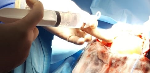
Case presentation: A 5-year-old boy from a rural area with no pathological history was admitted to pediatric emergency for sudden decreased level of consciousness with right‑sided hemiparesis and intermittent vomiting. On Physical examinations, he was disoriented with bilateral papilledema and right‑sided hemiparesis. Brain CT scan revealed a large cystic lesion in the left fronto‑parietal region with mass effect. There was no contrast enhancement , suggestive of hydatid cyst. Radiological investigations of thorax and abdomen disclosed no evidence of hydatid disease. The patient underwent craniotomy and the lesion was removed by irrigating saline between cyst wall and brain interface. Pathological examination confirmed hydatid cysts. Immediate improvement of the symptoms was observed and the patient was discharged on albendazole for 3 months.
Conclusion: The incidence of primary hydatid cyst of brain is very rare. The CT and MRI are the best diagnostic investigation and surgery extirpation of the intact cyst is the treatment of choice, resulting in complete recovery.

